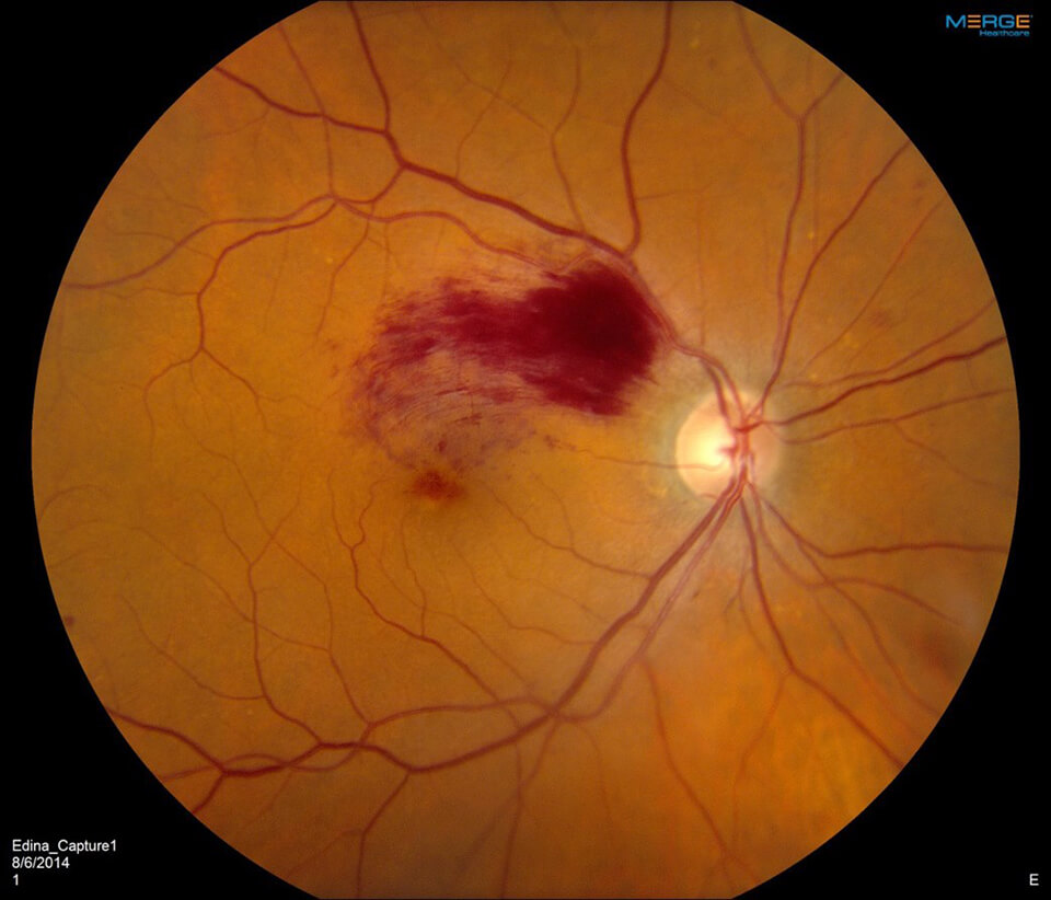
Branch Retinal Vein Occlusion
What Is Branch Retinal Vein Occlusion (BRVO)?
From reading to driving to perceiving color, much of our vision is made possible by the retina – a thin layer of photosensitive tissue that lines the inside of the back of the eye and connects to the brain through the optic nerve. Containing hundreds of millions of nerve cells, the retina acts similar to film in a camera by receiving light and converting them into neural signals that are sent to the brain, where they are interpreted and transformed into an image. The central region of the retina is known as the macula, which is the primary component responsible for central vision. Our ability to do everyday things such as seeing in fine detail, reading, or recognizing faces is highly dependent on the macula’s function.
For all of this to be possible, the retina and macula require a steady flow of blood and oxygen. As such, the retina has its own vasculature system consisting of arteries, veins, and branches. When one of these branches becomes obstructed, it is known as a branch retinal vein occlusion (BRVO). In some cases, BRVO can lead to loss of vision as well as other complications. BRVO affects both men and women equally and is typically seen in patients aged 50 or older. However, BRVO can also occur in younger patients.
Risk Factors of Untreated BRVO
There are several risk factors associated with the BRVO, including:
- Atherosclerosis, a cardiovascular disease in which cholesterol plaque builds up in the artery walls
- Smoking
- High blood pressure, or hypertension
- A history of strokes
- Heart or artery diseases
- Glaucoma
- High blood lipid levels
- Blood clotting issues
- Inflammatory conditions
- Infectious diseases
Symptoms BRVO

In BRVO, vision typically is impacted by blood and fluid seeping into the retina. In many cases, the blood and fluids may reabsorb slowly over a period of time, but it can take several months. Symptoms are generally determined by where the BRVO has occurred, with the most common symptoms being distorted peripheral vision or blurry vision. In some cases, there are no symptoms.
Aside from the symptoms, BRVO can also lead to complications that can have a significant impact on vision. These complications include:
- Swelling and fluid accumulation in the macula, also known as macular edema
- Abnormal blood vessel growth, also known as neovascularization
- Reduced blood flow to the macula, also known as macular ischemia
Diagnosing Branch Retinal Vein Occlusion
In order to properly diagnose BRVO, several specialized tests may be required, including:
- Fluorescein angiography, an imaging method that uses specialized dye to highlight blood vessels in the eye
- Ocular coherence tomography (OCT), an imaging method that uses infrared light to take high-resolution images of the retina
- Color fundus photography, which captures high-definition color images of the inside of the eye
Branch Retinal Vein Occlusion Treatment Options
Because BRVO cannot currently be cured, many doctors’ first step in treatment is to determine and manage any risk factors that can cause or exacerbate BRVO.
Outside of preventative care, doctors may also rely on laser therapy to help manage the symptoms of BRVO. According to the “Branch Vein Occlusion Study,” researchers found that laser coagulation could effectively improve visual prognosis in patients with BRVO after three years of follow-up. It is worth noting that multiple laser treatments are sometimes required to achieve the desired results.
Anti-vascular endothelial growth factor (anti-VEGF) medications such as Avastin, Lucentis, and Eylea are also commonly used in cases of BRVO where neovascularization or macular edema is present. These medications are injected directly into the eye and help to inhibit abnormal blood vessels from growing. Intravitreal steroid injections are also sometimes used.
However, the usage of steroids can sometimes lead to increased intraocular pressure in some patients, especially patients who have been diagnosed with glaucoma. While painless, this increased pressure must be monitored, as it can lead to irreversible vision loss. After receiving a comprehensive evaluation from a primary care provider, aspirin therapy may be recommended.
Retinal Neovascularization
Another serious potential problem in BRVO is that of retinal neovascularization. In advanced cases, abnormal blood vessels grow from the retina into the vitreous gel of the eye. Since these vessels are very fragile, this can lead to major bleeding (vitreous hemorrhage) and scar tissue formation. Consequently, this will cause floaters and an immediate, painless loss of vision. Laser photocoagulation treatment to the peripheral retina (pan-retinal photocoagulation) is very helpful in this situation. Laser treatment usually results in stabilization or even regression of the blood vessel growth. The bleeding will sometimes clear on its own, but if it does not, then an operation to remove the blood and the vitreous gel, called a vitrectomy, is sometimes performed. In severe cases of abnormal blood vessel growth, the retina may be pulled away from the wall of the eye (tractional retinal detachment), and this may require surgical repair.
Schedule a Consultation
To learn more about diabetic retinopathy, please call (855) 515-2020 to schedule a consultation.
We are proud to serve the communities of Minneapolis/St Paul, Brainerd, Duluth, Glencoe, Mankato, St. Cloud, and more.

